
What Happens When Leads Are Mispositioned?
A misplaced lead can:
- Simulate ST-segment elevation
- Mask arrhythmias
- Produce abnormal Q waves
These errors can result in:
- Unnecessary cardiac interventions
- Delayed treatments
- Patient harm
Types of Lead Systems and Correct Positioning
3-Lead System:
Used in basic monitoring:
- RA: Right upper chest
- LA: Left upper chest
- LL: Left lower abdomen
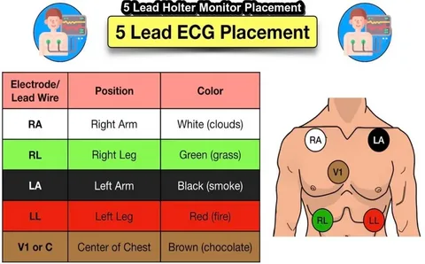
5-Lead System:
Standard in ICU and telemetry:
- RA: Below right clavicle
- LA: Below left clavicle
- RL: Lower right abdomen
- LL: Lower left abdomen
- V: Precordial position (depends on focus)
12-Lead System:
Vital for diagnostics:
- 6 limb leads (I, II, III, aVR, aVL, aVF)
- 6 chest leads (V1–V6)
Quick Visual Landmarks
- Midclavicular line for V4
- Anterior axillary line for V5
- Intercostal spaces for V1–V2
- Centered between ribs, never on top
Clinician’s Checklist
- ✅ Always use fresh electrodes
- ✅ Document any nonstandard placements
- ✅ Cross-check lead names and body sites
- ✅ Avoid artifact interference (e.g., chest hair, lotions)
Cardiac diagnostics rely heavily on accurate ECG readings. Yet, this accuracy hinges on something deceptively simple: the positioning of ECG leads. Whether you’re dealing with a basic 3-lead system or a comprehensive 12-lead diagnostic setup, placement precision matters more than you might think.
Wrap-Up
Mastering the positioning of ECG leads is a critical skill in modern medicine. Small placement errors can result in major clinical misjudgments. For reliable supplies and ECG education materials, medical professionals count on THE BIOMED GUYS.
